Saturday, January 30, 2010

Morbid Anatomy Presents at Observatory: "In the Henry Darger Archive," Illustrated Lecture by Jaimy Mann, 2009 Darger Study Center Research Fellow


Next Friday, February 5th, Morbid Anatomy and Observatory are proud to present an illustrated lecture about reclusive outsider artist Henry Darger by 2009 Darger Study Center Research Fellow Jaimy Mann.

Full details follow. Hope very much to see you there!
Morbid Anatomy Presents at Observatory
"In the Henry Darger Archive: From Rebellious Transsexual Child Slaves in Oz to Korean and Vietnamese Orphans"
An illustrated lecture by Jaimy Mann,, 2009 Henry Darger Study Center Research Fellow
Friday, February 5th
8:00 PM
$5

Outsider artist Henry Darger (1892-1973) is commonly remembered as a reclusive janitor who spent his life secretly creating a richly realized, bizarre and perverse fantasy world discovered only upon his death. While most who have considered Darger have seen his work as a spontaneous act of solitary and warped imagination, in tonight’s lecture, Darger scholar Jaimy Mann will reexamine the accepted narrative by discussing Darger’s work within the larger context of American cultural history and Darger’s personal biography. She will also introduce us to the holdings of the Henry Darger Archive held by the American Folk Art Museum in New York — where she conducted research as the 2009 Henry Darger Study Center Research Fellow — and give advice on how interested parties can gain access.

Contrary to popularly held beliefs, Henry Darger’s work–Mann argues– was not created in the vacuum of one cloistered man’s mind. While many of his paintings depict naked and brutalized child slaves and transexual transformation, L. Frank Baum’s phenomenally popular Oz books–which Darger read and collected– explored many similar topics normally taboo in the mainstream of American culture. “In the Realms of the Unreal,” Darger’s 15,000 page epic, draws from and inter-textualizes Oz, as well as the larger cultural obsessions with white slavery, seen, for example, in the passage of the “White Slave Traffic Act” in 1910. Further, Darger was personally affected by the instability and vulnerability that marks the existence of displaced children. When his mother died in childbirth, his baby sister was put up for adoption, and while in the home for feeble-minded children, Darger himself was almost adopted. He also desperately worked to adopt a child for decades as an adult, and in the archive, Mann investigated some rarely-exhibited collages (which will be on view at the American Folk Art Museum in 2010) that make use of photographs of Korean and Vietnamese children culled from newspapers. When looked at in this larger context of American cultural history, Mann believes that Darger is a key figure for understanding the notion and draw of “cuteness” in the United States, and will discuss how this notion is united with violence, race, and adoption.

Jaimy Mann is a Ph.D. candidate at the University of Florida, specializing in Children’s Literature and Culture and Gender Studies. She is based in Los Angeles, where she is raising her two-year-old daughter Querelle Magdalena while completing her dissertation “The Transnational Transracial Politics of Cute and Kawaii.” She was the 2009 Henry Darger Study Center Research Fellowship at the American Folk Art Museum in New York, where she had unlimited access to Darger’s books, paintings, and personal collection of newspaper clippings, collages, letters, religious items, and various other ephemera, which are housed in the museum archive. For more, please see http://jaimym.pixelfix.com
You can find out more by clicking here. You can get directions by clicking here. You can find out more about Observatory here, join our mailing list by clicking here, and join us on Facebook by clicking here.

Friday, January 29, 2010

"The Museum of Criminal Anthropology Cesare Lombroso"


Alessandro Molinengo--purveyor of Nautilus Scientific Antiques and Old Oddities of Turin, Italy (my favorite shop I've not yet been to!)--just emailed me about the newly re-opened "Museum of Criminal Anthropology Cesare Lombroso" at the University of Turin. Following is more about this amazing sounding museum, originally established in 1876 by infamous criminologist and physiognomist Cesare Lombroso, from the Nautilus website:
The Museum of Criminal Anthropology, dedicated to Cesare Lombroso, has reopened after years of restoration and access to specialist researchers only. The institution was founded by Lombroso in 1898 under the name "the Museum of Psychiatry and Criminology," documenting his beliefs and research into detecting criminality through physiognomy.

The 400 skulls in his collection, including one belonging to the brigand Giuseppe Villella, were used by Lombroso to develop his theory of the "median occipital fossa," a cranial anomaly that he believed contributed to deviant behavior.

On show are drawings, photos, criminal evidence, anatomical sections of "madmen and criminals" and work produced by criminals in the last century. The exhibits also include the Gallows of Turin, which were in use until the city's final hanging in 1865 and the possessions of a man known as White Stag, a renowned impostor who convinced Europe he was a great Native American chief. "But it is not a museum of horrors," insisted Giacomo Giacobini, coordinator of the "Museum of Man" project that the Lombroso collection will be part of. Rather, the museum is intended to recall positivistic era in science, in which Turin played a key role, starting with Cesare Lombroso's work.

The creation of the museum collections involved extensive interdisciplinary research by Lombroso in the fields of criminology, anatomy, psychiatry,psychology, sociology, ethnology, anthropology, linguistics, law, fine arts and medicine.

Lombroso's own head is also on display, a century down the line, perfectly preserved in a glass chamber.
Yet another reason to return to Italy! I hope very much to have the opportunity to visit this museum sooner rather than later, and the fabulous looking Nautilus Antiques as well!

All above images are drawn from the Nautilus Antiques write-up of the museum; to see many more images, and to learn much more about the museum, you can visit this original post by clicking here. You can visit the official "Museum of Criminal Anthropology Cesare Lombroso" website, and take a virtual tour of some of the artifacts, by clicking here. To check out the incredible Nautilus website (and see why I love it so!), click here. To find out more about Cesare Lombroso, click here. To read an earlier related Morbid Anatomy post, click here.

Philadelphia Medical Film Symposium Recap


I am still processing the many pleasures and terrors that comprised last week's Medical Film Symposium. I left the symposium completely exhausted, dreaming of bloodily sliced corneas and feeling that I might never need to see another surgical or venereal disease film ever again. But, on the other hand, I was much excited and stimulated by many things I saw or learned and the many fascinating people I had the opportunity to meet.

Highlights of the symposium included (but were not limited to):
  • Friday night's experimental film fest in the oldest operating theatre in the U.S., which I liken to being immersed in a kaleidoscopic, phantasmagoric fun house of medical horrors (but in a good way! See above images 2-6).

  • Michael Sappol's meta-lecture on the ethically-fraught pleasures of filming, collecting and viewing medical films paired with a screening of terrible and beautiful silent films from the National Library of Medicine (stay tuned for the opportunity to see this lecture at Observatory in Brooklyn! Click here to get on the mailing list and thus be alerted).

  • Oliver Gayken's lecture on wonder and science in early popular science films.

  • Getting to spend an entire day in the Mütter Museum's elegant and wonderful event space!

  • Saturday night's "Medical Film Cabinet of Curiosities" curated by Skip Elsheimer & Jay Schwartz, which gave all of us shell-shocked attendees the opportunity to laugh again, and made me long for the permissive and Utopian 1970s with the screening of the charmingly and innocently explicit school health film entitled "Achieving Sexual Maturity(1973)--completely unthinkable in today's social climate, with its nudity and celebration of youth sensuality, including on screen erections and masturbation--followed by a surrealistically charming school film demystifying a visit to the school nurse called "Just Awful" (1972).

  • The opportunity to take in the really wonderful Jan van Riemsdyk (aka van Rymsdyk) pastel exhibition outside the old operating theatre, which I highly recommend you check out if you are able before it closes in December 2010 (see bottom 2 photos; more on that here).

  • The grilled grapefruit at Reading Terminal's Down Home Diner!
All in all, although emotionally draining and a tad exhausting, the 2010 Medical Film Symposium was a wonderful experience, well organized and programmed, and I hope that the conference organizers have plans for a sequel in the near future.

You can find out more on the symposium by clicking here and here. You can find out about the Jan van Riemsdyk by clicking here.

Very special thanks to official symposium photographer Michelle Enemark (Observatory cohort and Curious Expeditions co-author and photographer) for the use of most of the above photos, and to conference organizers Dwight Swanson and Joanna Poses, for putting together such an inspired weekend and for giving me role of "official blogger."

Thursday, January 28, 2010

"Le Dernier Portrait," Morbid Anatomy Library Acquisition 1/26/10


The purpose of the exhibition is to evoke a practice of the past: portraying a deceased person, on their deathbed of in their coffin. This "last portrait" - death mask, painting, drawing or photograph - remained in the narrow circle of relatives and friends, but, in the case of famous personalities, it could be widely circulated in public. This practice, extremely common in Western countries in the nineteenth century and until the first half of the twentieth century, is today fast disappearing, or at least it remains strictly within the boundaries of the private sphere.

The exhibition gathers together pieces that are difficult to comment as they are linked to codes and rites now foreign to contemporary culture.

--The Musée d'Orsay website for the exhibition "Le Dernier Portrait"
The day before yesterday I visited the Burns Archive in New York for the first time. Stanley Burns' collection was really quite amazing, and I had a wonderful time viewing the incredible and impressive assemblage of photographs, books, and memorial artworks that surrounded him in his bustling brownstone home. I was also quite lucky to find that he had a few extra copies of a book I'd been coveting for sometime--Le Dernier Portrait, the catalog to an exhibition of the same name held at the Musée d'Orsay about 2 years ago--and was willing to sell me a copy for inclusion in the Morbid Anatomy Library.

The book, which translates to "The Last Portrait," explores the art and history of memorial and sickbed portraiture, touching on such portraiture in the fine arts, including examples by Munch, Gauguin, Seurat, Ensor, and Monet; 19th century memorial photography (featuring a selection of images from Burns' Collection, as featured in his incredible Sleeping Beauty: Memorial Photography in America); memorial tomb sculpture; news reportage; and the death mask.

This beautiful book now resides at the Morbid Anatomy Library; please feel free to come by and spend some time with it. You can find out more about the library by clicking here. For more on the Burns Archive, click here. For more on the book Le Dernier Portrait, click here. To find out more about the exhibition which inspired the book, click here.

Wednesday, January 27, 2010

"The Slashed Beauty," Clemente Susini, circa 1790


The beauty of a woman is only skin-deep. If men could only see what is beneath the flesh and penetrate below the surface with eyes like the Boetian lynx, they would be nauseated just to look at women, for all this feminine charm is nothing but phlegm, blood, humours, gall. Just imagine all that is hidden in nostrils, throat and stomach… We are all repelled to touch vomit and ordure even with our fingertips. How then can we ever want to embrace what is merely a sack of rottenness?
--Abbot Odo of Cluny, 10th Century, as found in Marina Warner's Phantasmagoria
Image from Anatomical Theatre Exhibition; Caption: "The Slashed Beauty," La Specola (Museo di Storia Naturale), Wax model with human hair in rosewood and Venetian Glass case; Probably modeled by Clemente Susini (around 1790), Florence, Italy;" click here to see full exhibition. Text via Jenny Hood. To find out more about Marina Warner's wonderful book Phantamagoria, click here.

Tuesday, January 26, 2010

"L'anatomie du corps humain: Avec ses maladies, & les remedes pour les guerir," Saint-Hilaire, 1684


You can browse the whole book by clicking here.

Monday, January 25, 2010

Happy "Blue Monday," The Most Depressing Day of the Year


As the Monday that begins the last full week in January, today has been accredited as "the most depressing day of the year." If this is the case, this year, so called 'Blue Monday' falls upon a rather fitting anniversary, as on the 25th January 1640, Robert Burton, author of The Anatomy of Melancholy died.
I have never heard of this phenomenon before, but I certainly believe it, and the weather in NYC today is definitely doing its part to make the prophecy seen quite true.

Read the full post on the Wellcome Library blog by clicking here. Image: "Melancholy," Domenico Feti, 1620, Musée du Louvre, Paris, found here.

Caterina de Julianis (1695-1742), Student of Gaetano Giulio Zumbo (1656-1701)


I have just made the happy discovery of the work of Caterina de Julianis (1695-1742), Neapolitan nun, master waxworker, and student of the Sicilian Abbot cum master waxworker Gaetano Giulio Zumbo (1656-1701). Zumbo was infamous in his time for his miniature allegorical waxworks depicting humans in the throes of death, disease, and decomposition; these artworks were found revolting by most, but beloved by a few, among them the Marquis de Sade. Zumbo is also famously known as the grandfather of the wax anatomical modeling tradition, as discussed in this recent blog post.

Julianis' work is so similar to that of her master--featuring waxwork miniatures, memento mori imagery and themes, and masterfully rendered macabre images of death and disease--that her work is often wrongly attributed to the better-known Zumbo; sometimes true ownership cannot be determined. Case in point: the top image is attributed on Victoria and Albert Museum website to both Julianis and Zumbo. The bottom image, from the same source, is entitled "Time and Death" and is attributed soley to Julianis, with a mention that it was, until recently, attributed to Zumbo. The full caption reads:
Time and Death, before 1727
Relief, Italy (probably Naples, made), Coloured and moulded wax
Purchased by the V&A under the bequest of Dr W.L. Hildburgh

The scene is set in a crumbling graveyard, with the winged figure of Father Time seated on the left pointing to a clock, while a half-draped emaciated figure of a smiling beggar, seated on the other side of the clock, solicits alms; a papal tiara lies at his feet. One small discoloured and decaying corpse lies in front of Time, while another corpse with entrails revealed lies beside him, surrounded by rats, snakes and skulls. A dead youth is stretched out on the right, while on the extreme right the crowned skeletal figure of Death holding a spear looks on. Ivy trails over the surrounding stonework; the sloping ground gives a sense of theatricality to the whole. The painted background depicts decaying funerary monuments.

This highly realistic and dramatic wax tableau was a memento mori, intended to inspire thoughts on mortality. Until recently it was attributed to the wax sculptor Gaetano Giulio Zumbo or Zummo (1656-1701), but it has now been convincingly reassigned to Caterina de Julianis. This artist was a Neapolitan nun who specialized in wax modelling. The piece was inspired by Zumbo's works, and the dead youth was in fact based on a figure of a dead bare-breasted woman in one of his wax compositions; because the present work was intended for a church this figure was transformed into a male subject. Coloured wax was the ideal medium for such morbidly realistic scenes, and the artist has been able to convey with astonishing illusionism the textures of stone, flesh and drapery. Wax figures could be formed from moulds, as well as modelled, and so copies and variations of compositions were easily made. A closely similar composition known to be by Caterina de Julianis is in the Chiesa dell'Immacolata in Catanzaro, previously in Bishop Emmanuel Spinelli's palace, and dating from before 1727.
It appears that one or both of these reliefs were featured in a recent exhibition at the V and A entitled "Baroque 1620-1800: Style in the Age of Magnificence." You can visit the online exhibition by clicking here; you can order a copy of the catalog (as I just did!) by clicking here. Both images from the V and A website; more on both objects here; click on images to see larger, finer image.

Addendum:
If the work of Zumbo is of interest, it has just come to my attention that one of Zumbo's recently restored artworks--described as "a gruesome scene showing a group of decomposing syphilis victims" (see below)

is on view for a limited time (until January 31st) at Florence's Museo dell'Opificio. For more on the piece and the exhibition, click here. And if these sorts of models hold some interest for you, you might also want to check out Daniel Neuberger's "Allegory on the Death of the Emperor Ferdinand" of 1657, which can be seen at the Ecclesiastical Treasury in Vienna and was featured in a recent MA post; click here to see it and find out more.

Sunday, January 24, 2010

Conjoined Twins, William Macewen, Date Unknown


From the fantastic Anatomy Acts exhibition, Object Guide No.151. Caption reads: "Conjoined Twins, date unknown, William Macewen, Courtesy of the Royal College of Surgeons of Edinburgh, LGR.

You can visit the entire online exhibition by clicking here; you can order the exhibition catalog by clicking here, or come by the Morbid Anatomy Library to spend some time with our own well-worn resident copy. Click in image to see in more detail.

Image via Rapeblossom.

Friday, January 22, 2010

Philadelphia Medical Film Symposium Weekend!


Today I am busing off for the weekend, to join the already-in-progress Philadelphia medical film symposium for a very exciting Friday night screening in the Pennsylvania Hospital's operating theatre, the oldest of its kind in the United States! Am also very much looking forward to tomorrow's day-long film symposium at the Mütter Museum, followed by Saturday night's "Medical Film Cabinet of Curiosities."

If you haven't registered for this amazing looking conference, do not despair; you can still attend screenings of your choice and pay on an event-by-event basis.

Hope very much to see you there! More on the details and locations of this event here.

Image, which I could swear I also saw in the original Wellcome Medicine Man Exhibit, via Rapeblossom.

Wednesday, January 20, 2010

Announcing a New Virtual Museum Dedicated to Frederik Ruysch (1638-1731): Anatomical Artist, Museologist, Morbid Anatomy Patron Saint!


There are many great artists of medical preparations, but perhaps my very favorite, and probably the most bizarre and fascinating to the modern eye, is Dutch anatomist, artist, preparator, and early museologist Frederik Ruysch. I have just been alerted to the launch of a new virtual museum dedicated to the life, art and history of this great anatomical artist. Following is a brief introduction to Ruysch's life and work; if you find this of interest, I urge you to visit this new virtual museum, which promises--once the English version becomes operative--to become a definitive resource for all things Ruysch.

Frederik Ruysch
(1638-1731) (pictured above, top, dissecting a child) was a true artist of human remains, his works being referred to in his time as "‘Rembrandts of anatomical preparation'" [1]. A high-ranking doctor in Amsterdam, Ruysch was famed far and wide for his uncannily life-like and imaginative preparations, and he used his access as "chief instructor of midwives and 'legal doctor' to the court" [2] to legally obtain scores of cadavers with which to create memorable preparations, including fanciful allegorical tableaux composed of fetal skeletons and other human body parts (as seen above, 2, 3 and 4). As Steven Jay Gould explains in the book Finders, Keepers: Eight Collectors, his excellent collaboration with photographer Rosamond Purcell
:
Ruysch made about a dozen tableaux, constructed of human fetal skeletons with backgrounds of other body parts, on allegorical themes of death and the transiency of life...Ruysch built the 'geological' landscapes of these tableaux from gallstones and kidneystones, and 'botanical' backgrounds from injected and hardened major veins and arteries for "trees," and more ramified tissue of lungs and smaller vessels for 'bushes' and 'grass.'

The fetal skeletons, several per tableau, were ornamented with symbols of death and short life--hands may hold mayflies (which live but a day in their adult state); skulls bemoan their fate by weeping into 'handkerchiefs' made of elegantly injected mesentery or brain meninges; 'snakes' and 'worms,' symbols of corruption made of intestine, wind around pelvis and rib cage.

Quotations and moral exhortations, emphasizing the brevity of life and the vanity of earthly riches, festooned the compositions. One fetal skeleton holding a string of pearls in its hand proclaims, 'Why should I long for the things of this world?' Another, playing a violin with a bow made of a dried artery, sings, 'Ah fate, ah bitter fate.' [3]
In addition to the spectacular memento mori-themed tableaux detailed above, Ruysch was also renowned for his incredibly life-like and enticingly imaginative wet preparations (above, 9, 10 and 11). To create these extraordinary specimens, Ruysch--using self-developed secret techniques--injected specimens with wax impregnated with pigment and other additives to solve the color-loss issues endemic to wet specimens. With the help of his daughter--still-life artist Rachel Ruysch--he would adorn these specimens with lace and clothing (sometimes even turbans! See 10 down) to hide "unfinished" areas (ie. cuts in the flesh, dissection marks) and add a note of delicacy, grace, and elegance to the whole; he would also often replace native eyes with eyes of glass to complete the illusion of life. The finished work would be immersed in his secret formula of spirits, black pepper, and other additives in a suitable presentation jar, with embellishments sometimes added atop the final piece (see 8 down). The results, as seen in the specimens above, were startlingly lifelike, and still--after many centuries!- have an uncanny, rosy beauty very unlike the dull and pallid specimens seen so often at medical museums.

Rusych was a collector and showman as well as artist and anatomist; to showcase his vast--
he produced over 2,000 human preparations in the years 1665-1717 alone! [4] --and "spectacular collections of 'Anatomical Treasures'," [5] Rusych established his own cabinet of curiosities, a private museum visited by medics and philosophers, as well as members of the aristocracy and royalty. Here, one could see not only his fantastic tableaux, but also "body part specimens in glass jars, baby skeletons, and preserved organs ... alongside exotic birds, butterflies and plants." [6] Ruysch published several lavishly illustrated guides to his incredible cabinet; 8th down, you can see an allegorical view of his museum as depicted in a frontispiece to one of these guides, Thesaurus animalium primus = Het eerste cabinet der dieren, published in 1710.

One visitor to the museum--and one of Ruysch's greatest admirers during his lifetime--was Czar Peter the Great of Russia, who in 1717 purchased the entire cabinet of Dr. Ruysch for 30,000 Dutch guilders, an astronomical fee at the time [7]. He had the entire collection shipped over to St. Petersburg--along with the collection of Amsterdam-based collector and scientist Albertus Seba--
to form the basis for the Academy of Sciences of Russia's first public museum, the Kunstkammer. [8]

Very sadly, none of Ruysch's astonishing tableaux are known to exist any longer, and are only known to us via illustrations from books of the time. Many of Ruysch's wet preparations, however, can still be viewed in collections such as the St. Petersburg Kunstkammer (which has 916 of them), Museum Bleulandinum in Utrecht, and the Anatomisch Museum LUMC in Leiden.

For people interested in seeing more of Ruysch's amazing work and learning more about the man and the collection, I suggest you visit the new online museum devoted to the man, his art and craft, and his collection. Entitled "The Anatomical Preparations of Frederik Ruysch," the site is organized by a Ruysch Research group formed between the St. Petersburg Kunstkamera the the University of Amsterdam. Although at the moment it is, unfortunately, available only in Dutch, it will eventually have operative English and Russian versions (more on that when these versions launch), and promises to be a great resource, with information about the work, history, collections, and techniques of Frederik Ruysch, and, incredibly, featuring a gallery linking many of the surviving specimens to the artist/anatomist's original descriptions of the objects--a rare museological feat!

You can visit the new virtual Ruysch Museum website by clicking here. To find out more about museums that still house Ruysch specimens, you can visit these sites: St. Petersburg Kunstkammer, Museum Bleulandinum, and the Anatomisch Museum LUMC in Leiden. For much more information, please visit the citations in the bibliography for this post, which you will find listed below. Another great resource is Steven Jay Gould and Rosamond Purcell's Finders, Keepers: Eight Collectors, which you can find out more about (and order a copy of) by clicking here; you can also visit this book and others that feature images of or information about Ruysch's work in the Morbid Anatomy Library, which is open by appointment.

Bibliography for this post, listed alphabetically: The Anatomical Preparations of Frederik Ruysch, BibliOdyssey, The British Library, Christies Auction House, Dream Anatomy, The St. Petersburg Kunstkammer, Medisch Erfgoed, She-Philosopher, The Smithsonian, University of Amsterdam, Wikipedia: frederik_Ruysch, Wikipedia: Kunstkamera, The Zymoglyphic Museum.

Images, top to bottom:
  1. Anatomische les van Dr. Frederik Ruysch, 1683; Jan van Neck (ca. 1634/'35 - 1714), Sourced here
  2. A skeleton cries into a handkerchief, © The British Library Board, Sourced here
  3. Crying skeletons with violin, feathers and fly, © The British Library Board, Sourced here
  4. Sourced from the Zymoglyphic Museum
  5. Skeleton with puppets, © The British Library Board, Sourced here
  6. Body part puppets, © The British Library Board, Sourced here
  7. Frederik Ruysch, (1638-1731). Thesaurus anatomicus primus [-decimus]... Het eerste [-tiende] anatomisch cabinet... Amsterdam: Jansson-Waesberg (part I and X), 1721 and 1716, Joannes Wolters (parts II-IX), 1702-1715., Christie's Auction House, Sourced here
  8. Frederik Ruysch (1638–1731). Arrangement of wax-injected anatomy specimens, found here
  9. Frederik Ruysch, Thesaurus animalium primus = Het eerste cabinet der dieren, 1710, Sourced here
  10. Child's Head with Turkish Cap, attributed to Frederik Ruysch, Photo © Rosamond Purcell, Sourced here
  11. Één van de anatomische preparaten van Frederik Ruysch, collectie Universiteitsmuseum, Nijmegen, Sourced here
  12. Specimen of a child’s arm. Frederik Ruysch, Sourced here
  13. Skeleton of Siamese twins. Preparation of Frederik Ruysch, Sourced here

Sunday, January 17, 2010

The Trope of the Mysterious Floating Hands


The Trope of the Mysterious Floating Hands, as seen in a life-sized wax model of a cesarean section from the Musee Spitzner (top) and an early 20th Century Polish poster (bottom).

Image of Spitzner wax model from the book Voir-La Collection Spitzner, eds. Phillipe Blon and Stephen Bann [1998]; more images can be found here. This book and others of its ilk can be visited at The Morbid Anatomy Library, about which more can be found by clicking here. The poster, with text reading "Zabawka" or "Toy," is by Stefan Norblin from 1933. It was found on Smashing Magazine's excellent feature "The Legacy Of Polish Poster Design;" full post (with many, many more excellent images) here.

Thursday, January 14, 2010

Morbid Anatomy Library Open Hours Followed by Observatory Opening, Saturday January 16th


This Saturday January 16th, the Morbid Anatomy Library will be open to the interested public from 1 PM until 6 PM. Come to the library for stack perusal and artifact investigation, and stick around for the opening party of our next-door-neighbor and partner Observatory's newest exhibition, "Vision Quest: A Group Show of Neo-Shamanic Art, "curated by Phantasmaphile's Pam Grossman.

Hope to see you there!

More about the library here; directions here. more about the Vision Quest exhibition here.

Photo: Eric Harvey Brown, for Time Out New York

Harvey Cushing (1869-1939)


More here. Via Elettrogenica.

Wednesday, January 13, 2010

Afternoon Skull Examination at the Swiss Benedictine Abbey of Einsiedeln


Afternoon skull examination at the Swiss Benedictine Abbey of Einsiedeln.

Via the always amazing Wurzletod Tumblr, who found it here. It appears to have originally derived from the Klosterarchiv Einsiedeln, though that's all the information find; caption from Wurzeltod's original post. Click on image to see one larger and lovelier.

Tuesday, January 12, 2010

Dan Estabrook, Arcane Media Artworks


My friend and sometime Observatory Helpmeet Megan Fitzpatrick just brought my attention to the lovely and mysterious arcane-technique-utilizing contemporary artworks of Dan Estabrook; above are just a few of my favorites; you can see much more of his work by clicking here.

Thanks, Megan, for sending this my way!

Monday, January 11, 2010

Medical Film Symposium, January 20-23, Philadelphia, PA


The Medical Film Symposium will examine—through screenings, presentations and papers—the relationship between moving images and medical science. Medical films comprised one of the earliest film genres, but the vast majority of these films are unseen and unknown today...
I can hardly wait for the upcoming Medical Film Symposium, taking place later this month in Philadelphia, PA--January 20-23 to be exact. As far as I know, this ambitious event--featuring over a dozen academics, film-makers and film-historians screening and expounding on films that explore "the relationship between moving images and medical science"--is the first of its kind, and promises to be a really fascinating, compelling, and, at times, perhaps even disturbing event.

As if the stellar line-up (see below) and opportunity to see obscure, under-seen old medical films weren't enough of a draw, the spaces housing the symposium are nearly as alluring as the events themselves; the incomparable Mütter Museum (!!!) will be hosting Saturday's day long program, and Friday night's screening will take place within (yes, within!) the Pennsylvania Hospital Surgical Theatre, the oldest surgical theatre in the United States. As an added bonus, attendees of this screening will have the opportunity to take in the Jan van Rymsdyk pastel drawings on view at the Pennsylvania Hospital's medical library--as mentioned in this previous post--before and after the event.

Other highlights of the symposium include Saturday night's "Medical Film Cabinet of Curiosities" co-curated by the Secret Cinema and North Carolina's A/V Geeks, and friend-of-Morbid-Anatomy Michael Sappol's presentation "Difficult Subjects," in which he will screen some harrowing medical film clips from the collection of The National Library of Medicine and "think aloud about the cultural meaning, scientific uses, ethical issues that arose in the making and showing of such films in their first historical moment" as well as today.

Full conference text follows; its a bit wordy, but well worth reading. The deadline for registration is this Friday, the 15th of January; the conference cost is only $80 ($50 for students), which includes admission to all events, plus breakfast and lunch on Saturday. I will definitely be there, in my role as "official blogger" for the event, and hope to see you there, too. This looks seriously not-to-be-missed, and lets hope it is the first of many such events exploring seriously the interstices of art and medicine, death and culture.
Medical Film Symposium, January 20-23, 2010

Wednesday, January 20
7:00pm

Screening of A Man to Remember at International House
(presented by Nico de Klerk of the Nederlands Filmmuseum) [info]
Preceded by opening of Radiologic Images exhibit (begins at 6:00pm)

Thursday, January 21
7:00pm

Film screening at International House
(curated by Barbara Hammer) [info]

Friday, January 22
7:00pm: Film screening within the historic Pennsylvania Hospital Operating Theatre
(curated by Andrew Lampert and Greg Pierce, open to symposium attendees only)

Saturday, January 23
9:00am to 5:00pm
A full day of presentations at the Mütter Museum
(Philadelphia College of Physicians )
  1. "The Body Visible"
    R. Nick Bryan, University of Pennsylvania

    While mankind has always been driven by morbid curiosity to see inside its own body, medical practitioners have had a more urgent need to do so – their business lies there-in. The spatial complexity of the body demands imaging not only for diagnosis but for successful treatment. However, the unaided human visual system that depends on visible light cannot see below the skin. Prior to Rontgen’s discovery of x-rays in 1895, the interior of the body could be imaged only by cutting through the skin and ‘letting the light in.’ Unfortunately, until the late 19th Century, such invasive medical imaging was usually performed after or immediately prior to death. Despite an initially slow and crude start with ‘Rontgenography’, the eternal goal of real time, safe, non-invasive, detailed imaging of the living human body has come to dramatic fruition in the past decade. With modern CT, nuclear, MR and ultrasound scanners, vivid static as well as moving images of all major organ systems are now routinely performed, as will be illustrated by videos of, “My Body”, a self-exposé by the presenter.

  2. "Between Photography and Film: Early Uses of Medical Cinematography"
    Scott Curtis, Northwestern University

    From the beginning, medical researchers and physicians eagerly appropriated the new technology of motion pictures. For some, especially those interested in a more "scientific" approach to medicine, film represented an improvement upon and transformation of serial photography--that is, they regarded motion pictures as a series of still images. Others extended medical photography's more common use as documentary evidence to their application of cinema. Still others emphasized the spectacular and moving quality of the cinematic image in their promotion of film as an educational tool, often distinguishing it from photography. This presentation, then, will survey the professional perceptions and uses of medical cinematography in its first two decades and compare those uses to the functions, genres, and venues of nineteenth- and early-twentieth-century medical photography.

  3. "The Flow of Life: Moving Images of Magnified Blood"
    Oliver Gaycken, Temple University

    A staple of medical moving-image presentations was the spectacle of blood as seen under a microscope projected onto a screen. This talk will consider some examples of this tradition that range from nineteenth-century lantern lectures to reminiscences of researchers to the incorporation of this genre into the medical motion picture.

  4. "Complexities and Enigmas of Cinefluorography in the work of Dr. James Sibley Watson and Colleagues"
    Barbara Hammer (Independent filmmaker) and Patti Doyen (George Eastman House

    This presentation will explore through Watson et. al.'s text and images the discoveries and problems of the Rochester medical team that led to mechanical inventions that enabled views of the interior of the human body. The uses and abuses of the techniques will be highlighted as well as the artistic curiosities Watson pursued in spectacles that had no scientific purpose.

  5. "Telephone Operator, Camera-Operator: Laryngoscopy and High Speed Motion Pictures at Bell Labs"
    Mara Mills, University of Pennsylvania

    During the early twentieth century, telephone engineers became authorities on psychoacoustics and otolaryngology. In the interests of visualizing speech production and the movement of circuit components, they also made key contributions to high speed motion picture photography. This talk will survey the history of laryngoscopy through the 1940s, concluding with a few remarks about the nature of the "telephonic gaze."

  6. "Edgar Ulmer and the National Tuberculosis Association: Fighting Faith in the War Against TB"
    Devin Orgeron, North Carolina State University

    From the late 1930s through the early 1940s, well-known “B” movie director Edgar Ulmer (sometimes called the King of PRC) directed eight health shorts for the National Tuberculosis Association. A strain of fatal contamination runs though all of Ulmer’s work and is brilliantly, if oddly articulated in these tuberculosis films, many of which are aimed at specific American racial minorities and the inadequacies of their sometimes imported faith in the face of the disease. Along with their fit within Ulmer’s career, I hope to illustrate the role these films played in shaping 1930s/1940s notions of race, religion, and disease.

  7. “‘Spectacular Problems in Surgery’: Medical Motion Pictures at the American College of Surgeons”
    Kirsten Ostherr, Rice University

    Early in the twentieth century, the American College of Surgeons was a leading national force in the use of motion pictures for educational purposes. This movement encompassed all facets of the motion picture industry (ranging from education to entertainment), and established the ACS as a central institution in the history of cinema. Moreover, the ACS became an important vehicle for international medical education through motion pictures after World War II, and this aspect of ACS activities provides an important and unique perspective on the varied global uses of medical media in the postwar era. This presentation will address the medical motion pictures produced, reviewed, distributed, and exhibited by the ACS, from the late 1920s to the present. The talk will be based on research at the American College of Surgeons archive, which contains paper records related to a vast range of medical motion pictures. These films were primarily technical medical films produced by specialists for other specialists, as well as for medical student and resident training. Since the ACS films were concerned not only with medical education but also with the public image of the medical profession, this history serves a critical function in assessing the role of visual images in shaping the popular and specialist cultures of medicine throughout the twentieth century.

  8. "Difficult Subjects: Working with Films from the Collection of the National Library of Medicine"
    Michael Sappol, History of Medicine Division at the National Library of Medicine

    Historical medical film is notable for its representation and documentation of "difficult subjects"—the interior of the body, death, disgfigurement, radical medical intervention, infliction of pain on patients and research subjects, behavioral disturbance, venereal disease, emotional and physical distress, etc. Although publicly available, such films are rarely screened and, as a result, rarely studied. This presentation will screen a selection of these difficult films, explore their unique history, uses and abuses, effects on viewers, and the larger issues that they raise.

  9. “Research, education, and patient care: archival medical film collections at academic health institutions”
    Timothy Wisniewski, Alan Mason Chesney Medical Archives, Johns Hopkins University

    This presentation will focus on the institutional context of archival film collections produced within academic health centers, using the Johns Hopkins Medical Institutions as the primary example. The presentation will look at historical examples of centralized and decentralized models of film production at Johns Hopkins, and compare genres of medical film as produced for educational, clinical, or biomedical research purposes. Finally, the presentation will discuss the value of making these often unprocessed or restricted collections accessible for research and use by diverse groups of users.
Saturday, January 23
8:00 pm
A MEDICAL FILM CABINET OF CURIOSITIES
Film screening at Moore College of Art (curated by Skip Elsheimer & Jay Schwartz)
Admission: $7.00 [admission included with symposium registration]

Co-programmed by Jay Schwartz of the Secret Cinema and Skip Elsheimer of North Carolina's A/V Geeks, who will be present at the screening. This will be Skip's first return visit since he presented his popular program S IS FOR SISSY! at Moore, just over one year ago.

The program will include:
  • FEET AND POSTURE (1920s) - This reel, from the earliest era of 16mm educational films, aims to explain the physiology of feet and how to best take care of them. It demonstrates through x-rays how the well-dressed young flapper of the time often did not choose the best footwear. Made with the cooperation of M.I.T. and the American Posture League.

  • CELL WARS (1987) A lively introduction to immunology that shows kids how the body´s cells defend themselves against invading germs. Crazy-costumed actors and dazzling video effects demonstrate what happens after germs enter the body through a skinned knee.

  • CRYOEXTRACTION (195?) - A sales and demonstration film showing off the Thomas Cryopter--a device which resembles a power router, which is then shown in use for eye surgery.

  • COLDS AND FLU (1975) - Kids dressed in armor battle each other to seize control of a giant-mouthed castle.

  • ACHIEVING SEXUAL MATURITY (1973) - At a time when DEEP THROAT played in neighborhood cinemas alongside traditional Hollywood fare, educators struggled as to how to best meet increasingly rebellious high school and college students on their own terms. It was during this possibly unique moment in pop culture that ACHIEVING SEXUAL MATURITY was successfully sold to school districts around the country. Its use of graphic live photography of nude males and females to explain and illustrate sexual anatomy from conception to adulthood is today quite surprising.

  • NON-SYPHILITIC VENEREAL DISEASE (195?) - This short film made for the medical community--in still-stunning Kodachrome color -- details a variety of exotic venereal diseases, in close-up after horrifying close-up. This mainstay of Secret Cinema Halloween screenings is guaranteed to have audiences screaming in terror.

  • JUST AWFUL (1972) This film was made to help eradicate any fears children may have about visiting the school nurse.
SECRET CINEMA WEBSITE: http://www.thesecretcinema.com
For more information about the symposium, visit the conference website by clicking here; you can find out more about registration here. For more about symposium hosts the Mütter Museum and Pennsylvania Hospital Surgical Theatre, click here and here, respectively. Please feel free to contact me with any questions by clicking here.

Hope to see you there!

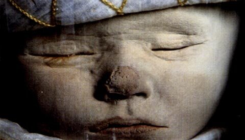
Images: From top: Still from SANCTUS, dir. Barbara Hammer, Courtesy of Barbara Hammer; X-rays by Dr. James Sibley Watson, Courtesy of Barbara Hammer; Maurice L. Blatt, Samuel J. Hoffman, & Maurice Schneider, “Rabies: Report of Twelve Cases, with a Discussion of Prophylaxis,” Journal of the American Medical Association 111 (1938): 688-91